Don't worry about the people in your past; There's a reason they didn't make it to your future.
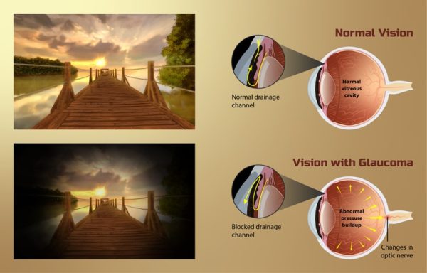
Glaucoma – Silent Thief of Sight
Glaucoma is an eye disorder that causes damage to the optic nerve that carries information from the eye to the brain. The increased pressure affects the optic nerve and may cause vision loss.
Two types of classification of glaucoma are open-angle (usually painless) and angle-closure or narrow-angle glaucoma (often associated with pain and redness of the eye). The “angle” in both cases refers to the drainage angle inside the eye that controls the outflow of the watery fluid that is continually being produced inside the eye.
Treatment:Glaucoma usually has few or no initial symptoms. By the time vision is affected, the damage is permanent. The early diagnosis is key for glaucoma. Progression of glaucoma can be slowed or halted with eye drops, laser treatments or surgery. Researchers has found out that, higher level of exercise appear to provide a long-term benefit of reducing the incidence of low ocular perfusion pressure which is a main factor of glaucoma. Glaucoma development can be reduced by not smoking, maintaining a healthy weight, and eating a varied and healthy diet.
Reference: http://www.allaboutvision.com/
Image credit: https://www.myupchar.com/en, CC BY-SA 4.0 <https://creativecommons.org/licenses/by-sa/4.0>, via Wikimedia Commons
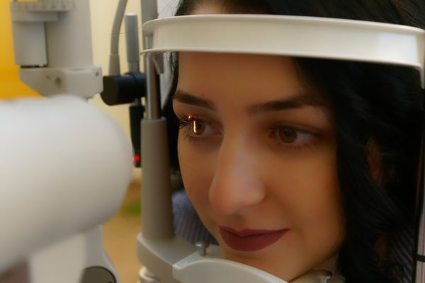
A cataract is clouding of eye lens (between iris and pupil) that results in blurry vision. With age, it progresses slowly. Cataract is the main cause of blindness in the world. People who are above 35 may develop cataract. The other causes of cataracts include conditions like diabetes, trauma, some medications, improper diet, smoking and exposure of eyes to excessive UV light.
Types of cataracts are:
1) A subcapsular cataract occurs at the back of the lens -people with diabetes and who take high amount of steroid medications develop this type of cataract
2) A nuclear cataract forms deep in the central zone of the lens associated with aging and
3) A cortical cataract is characterized by white, wedge-like opacities that start in the periphery of the lens and work their way to the center in a spoke-like fashion.
Treatment:Routine eye examine helps in determining cataract development. Treatments include eyeglasses, magnifying lenses, or surgery. Surgery is curative as the cloudy lens is removed and replaced with an artificial one.
Reference: http://www.allaboutvision.com/
Image credit: Image by Paul Diaconu from Pixabay (cc by 0)
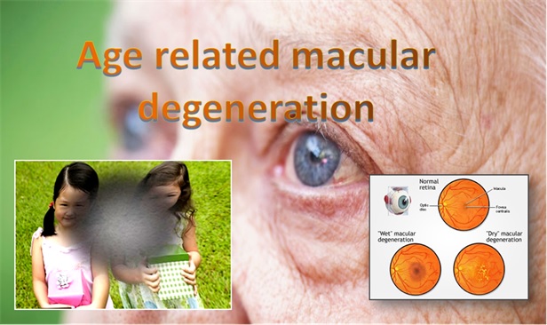
Age-related macular degeneration or macular degeneration is an eye disease with onset at any age, usually after age 60, that progressively destroys the macula- the central portion of the retina that helps with focus. It rarely causes total blindness as only the center of vision is affected. The health of the macula determines our ability to read, recognize faces, drive, watch television, use a computer, and perform any other visual task that requires us to see fine detail.
There are two types of macular degeneration: wet and dry. In wet AMD, abnormal blood vessels behind the retina start to grow, leaking blood and fluid, causing loss of central vision, which may occur quickly. In dry AMD, which is more common, the light-sensitive cells in the macula slowly break down causing central vision to diminish over time.
Treatment:Yellowish spots known as drusen begin to accumulate in and around the macula when there is dry AMD develops. There are no proper approved medications are available for this condition. However, nutritional supplements containing antioxidant vitamins and multivitamins that also contain lutein and zeaxanthin can reduce the risk of dry AMD progressing to sight-threatening wet AMD.
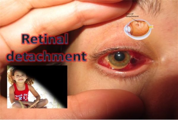
The retina is the light-sensitive layer of tissue that lines the inside of the eye and sends visual messages through the optic nerve to the brain. When the retina detaches, it is lifted or pulled from its normal position. If not promptly treated, retinal detachment can cause permanent vision loss. Retina detachments are often painless, and symptoms that may be noticed include perception of flashing lights, floaters, or a curtain drawn over your visual field.
Rhegmatogenous –The most common type where a tear or break in the retina allows fluid to get under the retina and separate it from the retinal pigment epithelium (RPE), the pigmented cell layer that nourishes the retina.
Tractiona – This less common where a scar tissue on the retina’s surface contracts and causes the retina to separate from the RPE.
Exudative – This is caused by retinal diseases such as inflammatory disorders and injury/trauma to the eye. Fluid leaks into the area underneath the retina, but there are no tears or breaks in the retina.
Treatment:Treatment for a detached retina involves surgery, mostly using lasers, that can improve vision affected by the retinal detachment. Small holes and tears are treated with laser surgery or a freeze treatment called cryopexy.
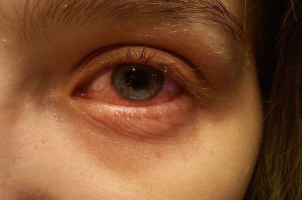
This is redness and inflammation of the clear tissue covering the eye and the inside of the eyelids (conjunctiva). Most cases infections caused by viruses or bacteria results in pink eye. The other factors -dry eyes from lack of tears or exposure to wind and sun, Chemicals, fumes, or smoke (chemical conjunctivitis) and allergies due to pollens, dust etc. During pink eye a crusty discharge may make it difficult to open the eyelids.
Treatment: Bacterial conjunctivitis can be treated with antibiotic drops or ointments prescribed by doctor. Most cases of infectious conjunctivitis are viral and do not need treatment with antibiotics. If there is crusty discharge then use a warm, wet compress and gently remove the crusting on the eyes. To reduce the spread of the infectious conjunctivitis, wash hands frequently, do not share eye drops, cosmetics, towels, or washcloths.
Image credit: Daemonanyndel, CC BY-SA 3.0 <https://creativecommons.org/licenses/by-sa/3.0>, via Wikimedia Commons
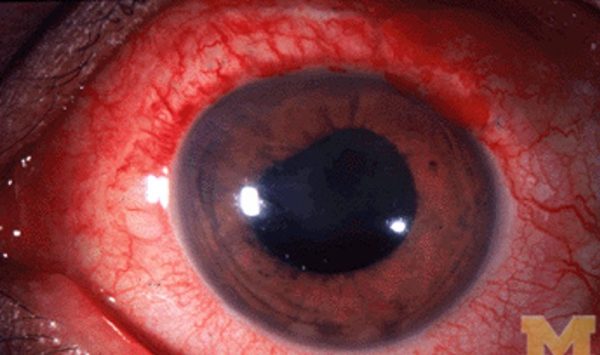
Uveitis is inflammation of the uvea, which is made up of the iris, ciliary body and choroid. Together, these form the middle layer of the eye between the retina and the sclera (white of the eye). Causes of uveitis include trauma or injury to the eye, infections, or rheumatologic or inflammatory diseases that affect other parts of the body. Uveitis may be associated with:
A virus, such as shingles, mumps or herpes simplex;
Systemic inflammatory diseases;
A result of injury to the eye; or
Rarely, a fungus, such as histoplasmosis or a parasite, such as toxoplasmosis.
The main symptom of uveitis is pain in the eyeball. The eye will look red (bloodshot) and you may notice blurred vision, light sensitivity, and spots in your vision.
Treatment: Treatment for uveitis depends on the cause. Uveitis is a serious eye condition that may scar the eye. It needs to be treated as soon as possible. Eye drops, especially corticosteroids and pupil dilators, can reduce inflammation and pain. If it is not treated, it may result in glaucoma, cataract, damage of retina and neovascularization.
Image credit: http://kellogg.umich.edu/theeyeshaveit/redeye/anterior_uveitis.html (CC by 3.0)
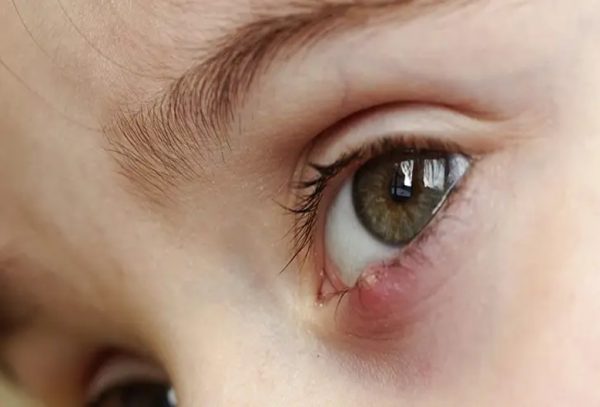
A sty (stye) is an infection of the oil gland at the base of an eyelash. It is a tender, painful red bump located at the base of an eyelash or inside the eyelid. It appears as a red, raised pimple on the edge of the eyelid. Symptoms of a sty can be pain, tenderness, redness, and swelling with a small pustule. The eyeball itself may feel irritated or as if something is scratching it due to the swelling of the eyelid.
Treatment for a sty includes warm compresses applied to the affected area for 10 minutes, up to six times daily. If the sty comes to a head and releases pus, it should be cleaned gently with soap and water. A sty should not be pressed or squeezed to facilitate drainage. If a sty persists for several days, a doctor may lance (drain) the infection under local anesthesia. If the sty is very large, painful, or affects your vision, see your doctor.
Chalazion (Eyelid Cyst):A chalazion (meibomian cyst, tarsal cyst, or conjunctival granuloma) is a lump in the upper or lower eyelid caused by obstruction and inflammation of an oil gland of the eyelid. Eyelid glands are called the meibomian glands The gland opening becomes clogged and the gland swells.
Treatment:Chalazia are treated with warm compresses, though in rare cases they may require antibiotics. If the chalazion becomes severe, causes changes in vision, or is persistent, it may be removed surgically.
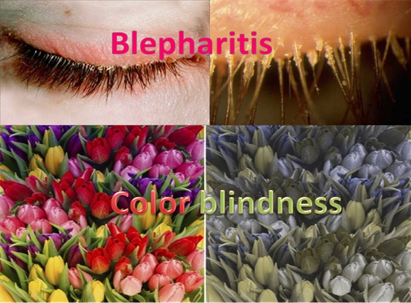
Blepharitis is inflammation of the eyelids. The inflammation can be found on the outer or inner eyelid. The condition can be difficult to manage because it tends to recur. Symptoms of blepharitis include burning, itching, swelling, flaky skin at the base of the lashes, crusting of the eyelids, blurred vision excessive tearing, itching, sensitivity to light. Other causes of blepharitis can be infections, or other skin conditions.
Treatment:Good eyelid hygiene, including frequent cleaning, light scrubbing, using a mixture of water and baby shampoo. Severe cases of blepharitis may require antibiotics or steroids.
Color Blindness:The colors we see are a result of how our eyes (and thus our brains) interpret different wavelengths of light. People with color blindness have difficulty seeing certain colors, usually reds, greens, and blues. Inherited color blindness is caused by abnormal photopigments. Most of the time this is genetic but it can also be caused by aging, disease, trauma to the eye, or certain medications. If the cause of the color blindness is genetic, the problem cannot be corrected but people may be trained to adapt to interpret color shades. Men are much more likely to be colorblind than women because the genes responsible for the most common, inherited color blindness are on the X chromosome. In cases where color blindness is acquired, it may be treatable.
Treatment: There is no cure for color blindness. However, people with red-green color blindness may be able to use a special set of lenses to help them perceive colors more accurately. These lenses can only be used outdoors under bright lighting conditions. Visual aids have also been developed to help people cope with color blindness. There are iPhone and iPad apps, for example, that help people with color blindness discriminate among colors.