


 Fusion
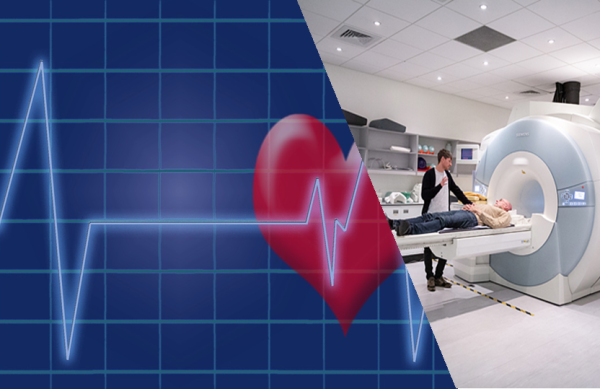
Electrocardiogram And Magnetic Resonance Imaging For Heart
3. Electrocardiogram (ECG or EKG): Measures the electrical activity of the heartbeat to provide two kinds of information. First, by measuring time intervals on the ECG, a doctor can determine how long the electrical wave takes to pass through your heart. Finding out how long a wave takes to travel from one part of the heart to the next shows if the electrical activity is normal, slow, fast or irregular.
Second, by measuring the amount of electrical activity passing through heart muscle, a cardiologist may be able to find out if parts of the heart are too large or overworked.
Reasons for the test:
- Monitor changes in heart rhythm
- Determine whether a heart attack has occurred
- Help predict if a heart attack is developing
4. Magnetic resonance imaging (MRI): Uses a magnetic field and radiofrequency waves to create detailed pictures of organs and structures inside your body. It can be used to examine your heart and blood vessels and to identify areas of the brain affected by stroke.
Reasons for the test:
- Assess heart structure
- Look for scar tissue within the heart muscle
- Assess the function of heart valves
Reference: https://healthblog.uofmhealth.org/
Image credit:
- https://www.maxpixel.net/Electrocardiogram-Ecg-Heartbeat-Pulse-Heart-1892826
- https://commons.wikimedia.org/wiki/File:190603_Functional_magnetic_resonance_imaging_at_the_Imperial_Centre_for_Psychedelic_Research.jpg : Thomas Angus, Imperial College London, CC BY-SA 4.0 , via Wikimedia Commons
Author: HealthyLife | Posted on: November 2, 2021
« Previous Article
Next Article »
« CT Scan And Exercise Cardiac Stress Test For Heart What are the tests that help us to determine our heart health? »
« CT Scan And Exercise Cardiac Stress Test For Heart What are the tests that help us to determine our heart health? »


Write a comment