


 Fusion
Retinal Detachment Symptoms, Precautions and Treatment
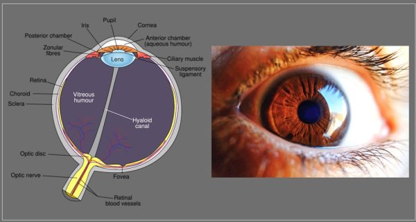
Retina is a light sensitive layer of tissue situated in the back of the eye. Retinal detachment is an eye issue that happens when the retina is pulled away from its position at the back of the eye. Retinal detachment is an emergency and need to immediate attention. If not treated on time, more detachment happens resulting in permanent loss of vision.
Symptoms of retinal detachment:
When there is a small detachment, a person may not notice any types of symptoms. If the detachment is more then following symptoms one might face:
- Small dark spots or blurred lines that float across the eyes. Small floaters in front of the eyes
- Flashes of light in one or both eyes
- Experiencing dark shadow on sides or in the middle of the vision field
Who is at risk of retinal detachment?
Some people with certain conditions are more at risk of retinal detachment. But it can also happen to anyone.
- People who have family with history of retinal detachment
- Person with serious eye injury
- An eye surgery like treatment to cataracts
- Diabetic condition called retinopathy where high blood sugar affects blood vessels of the retina
- Person with extreme nearsighted condition called myopia
- A person with posterior vitreous detachment where the gel like fluid in the center of eye pulls away
- Degeneration or thinning of retina
- Condition called retinoschisis where retina separates to two layers
- The most common causes is aging
- Tumors in the eye
- Regular inflammation inside the eye
- Rare eye disorder where abnormal development of blood vessels in retina – coats disease.
The three types of retinal detachment are:
Rhegmatogenous : When the retina has a tear or break, the gel-like fluid in the center called citreous can get behind retina and pushes retina away from the back of the eye.
Tractional: Tractional detachment is when scar tissue on retina pulls retina away from the back of the eye.
Exudative: When the fluid builds up behind the retina, but there aren’t any tears or breaks in your retina
Prevention of retinal detachment:
- When it is age related there is no way to prevent retinal detachment. But one can lower the risk of the retinal detachment by wearing safety goggles or by protecting eye with eye gear during daily activities.
- If you are falling in any of the risk factor, it is highly recommended to get your eyes examined. An ophthalmologist will dilate eye and exam the eyes for retinal detachment before it starts affecting the vision.
- If you suffer from eye trauma it is important to see the eye doctor. Trauma could be an accident to head, hitting eyes, chemicals reaction etc.
- If you start seeing floaters and black shadows in your vision, immediately make an appointment for eye test.
Tests for observing retinal detachment:
Apart from dilating and examining eyes, your doctor may ask you to go through detailed examination. An ultrasound or optical coherence tomography (OTC) will give details. These tests are painless and will help doctors to understand retina condition.
Treatment for retinal detachment:
Treatment for retinal detachment depends on how much retina is detached and which type of detachment a person is suffering from.
Eye surgery, laser treatment, freezing treatment or treatment to fix tears and reattach retina in the back of eye might be performed. Sometimes doctor might use more than one treatment.
- Eye surgery: If more retina is detachment a surgery is helpful in putting back retina in place.
- Freeze treatment or laser surgery: Also known as cryopexy is helpful in fixing tear or small hole in retina by using medical laser or freezing probe tears will be sealed.
If caught early and treated immediately retinal detachment can be taken care to get back the proper vision. If you see floaters, shadows or any symptoms of change in your vision contact your eye doctor today for further testing and protect your vision.
References:
Image credit: Image by Clker-Free-Vector-Images from Pixabay (cc by 0) & Image by Juraj Varga from Pixabay (cc by 0)
Author: Sumana Rao | Posted on: June 12, 2024
« Previous Article
Next Article »
« Canker Sores Symptoms And Prevention Lateral Hip Pain Gluteal Tendinopathy Causes, Symptoms And Treatment »
« Canker Sores Symptoms And Prevention Lateral Hip Pain Gluteal Tendinopathy Causes, Symptoms And Treatment »


Write a comment